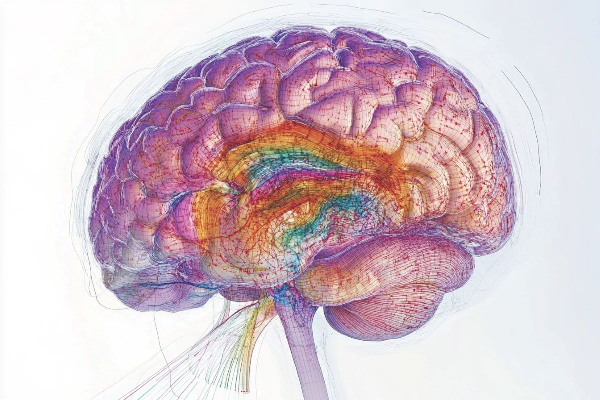
Summary: New research shows that not all brain cells age equally, with certain cells, such as those in the hypothalamus, experiencing more age-related genetic changes. These changes include reduced activity in neuronal circuitry genes and increased activity in immunity-related genes.
The findings provide a detailed map of age-sensitive brain regions, offering insights into how aging may influence brain disorders like Alzheimer’s. This research could guide the development of treatments targeting aging-related brain changes and neurodegenerative diseases.
Key Facts:
- Uneven Aging: Hypothalamic neurons and ventricle-lining cells show the greatest age-related genetic changes.
- Gene Activity Shifts: Aging reduces neuronal circuit genes but increases immunity-related genes.
- Therapeutic Potential: Mapping age-sensitive cells may inform treatments for aging-related brain diseases.
Source: NIH
Based on new brain mapping research funded by the National Institutes of Health (NIH), scientists have discovered that not all cell types in the brain age in the same way.
They found that some cells, such as a small group of hormone-controlling cells, may undergo more age-related changes in genetic activity than others.
The results, published in Nature, support the idea that some cells are more sensitive to the aging process and aging brain disorders than others.

“Aging is the most important risk factor for Alzheimer’s disease and many other devastating brain disorders. These results provide a highly detailed map for which brain cells may be most affected by aging,” said Richard J. Hodes, M.D., director of NIH’s National Institute on Aging.
“This new map may fundamentally alter the way scientists think about how aging affects the brain and also provide a guide for developing new treatments for aging-related brain diseases.”
Scientists used advanced genetic analysis tools to study individual cells in the brains of 2-month-old “young” and 18-month-old “aged” mice.
For each age, researchers analysed the genetic activity of a variety of cell types located in 16 different broad regions — constituting 35% of the total volume of a mouse brain.
Like previous studies, the initial results showed a decrease in the activity of genes associated with neuronal circuits.
These decreases were seen in neurons, the primary circuitry cells, as well as in “glial” cells called astrocytes and oligodendrocytes, which can support neural signaling by controlling neurotransmitter levels and electrically insulating nerve fibers.
In contrast, aging increased the activity of genes associated with the brain’s immunity and inflammatory systems, as well as brain blood vessel cells.
Further analysis helped spot which cell types may be the most sensitive to aging. For example, the results suggested that aging reduces the development of newborn neurons found in at least three different parts of the brain.
Previous studies have shown that some of these newborn neurons may play a role in the circuitry that controls some forms of learning and memory while others may help mice recognize different smells.
The cells that appeared to be the most sensitive to aging surround the third ventricle, a major pipeline that enables cerebrospinal fluid to pass through the hypothalamus.
Located at the base of the mouse brain, the hypothalamus produces hormones that can control the body’s basic needs, including temperature, heart rate, sleep, thirst, and hunger.
The results showed that cells lining the third ventricle and neighbouring neurons in the hypothalamus had the greatest changes in genetic activity with age, including increases in immunity genes and decreases in genes associated with neuronal circuitry.
The authors noted that these observations align with previous studies on several different animals that showed links between aging and body metabolism, including those on how intermittent fasting and other calorie restricting diets can increase life span.
Specifically, the age-sensitive neurons in the hypothalamus are known to produce feeding and energy-controlling hormones while the ventricle-lining cells control the passage of hormones and nutrients between the brain and the body.
More research is needed to examine the biological mechanisms underlying the findings, as well as search for any possible links to human health.
The project was led by Kelly Jin, Ph.D., Bosiljka Tasic, Ph.D., and Hongkui Zeng, Ph.D., from the Allen Institute for Brain Science, Seattle. The scientists used brain mapping tools — developed as part of the NIH’s Brain Research Through Advancing Innovative Neurotechnologies® (BRAIN) Initiative – Cell Census Network (BICCN) — to study more than 1.2 million brain cells, or about 1% of total brain cells, from young and aged mice.
“For years scientists studied the effects of aging on the brain mostly one cell at a time. Now, with innovative brain mapping tools – made possible by the NIH BRAIN Initiative – researchers can study how aging affects much of the whole brain,” said John Ngai, Ph.D., director, The BRAIN Initiative®.
“This study shows that examining the brain more globally can provide scientists with fresh insights on how the brain ages and how neurodegenerative diseases may disrupt normal aging activity.”
Funding: This study was funded by NIH grants R01AG066027 and U19MH114830.
About this aging and brain mapping research news
Author: Christopher Thomas
Source: NIH
Contact: Christopher Thomas – NIH
Image: The image is credited to Neuroscience News
Original Research: Open access.
“Brain-wide cell-type specific transcriptomic signatures of healthy aging in mice” by Kelly Jin et al. Nature
Abstract
Brain-wide cell-type specific transcriptomic signatures of healthy aging in mice
Biological ageing can be defined as a gradual loss of homeostasis across various aspects of molecular and cellular function. Mammalian brains consist of thousands of cell types, which may be differentially susceptible or resilient to ageing.
Here we present a comprehensive single-cell RNA sequencing dataset containing roughly 1.2 million high-quality single-cell transcriptomes of brain cells from young adult and aged mice of both sexes, from regions spanning the forebrain, midbrain and hindbrain.
High-resolution clustering of all cells results in 847 cell clusters and reveals at least 14 age-biased clusters that are mostly glial types.
At the broader cell subclass and supertype levels, we find age-associated gene expression signatures and provide a list of 2,449 unique differentially expressed genes (age-DE genes) for many neuronal and non-neuronal cell types.
Whereas most age-DE genes are unique to specific cell types, we observe common signatures with ageing across cell types, including a decrease in expression of genes related to neuronal structure and function in many neuron types, major astrocyte types and mature oligodendrocytes, and an increase in expression of genes related to immune function, antigen presentation, inflammation, and cell motility in immune cell types and some vascular cell types.
Finally, we observe that some of the cell types that demonstrate the greatest sensitivity to ageing are concentrated around the third ventricle in the hypothalamus, including tanycytes, ependymal cells, and certain neuron types in the arcuate nucleus, dorsomedial nucleus and paraventricular nucleus that express genes canonically related to energy homeostasis.
Many of these types demonstrate both a decrease in neuronal function and an increase in immune response.
These findings suggest that the third ventricle in the hypothalamus may be a hub for ageing in the mouse brain.
Overall, this study systematically delineates a dynamic landscape of cell-type-specific transcriptomic changes in the brain associated with normal ageing that will serve as a foundation for the investigation of functional changes in ageing and the interaction of ageing and disease.